
The shoulder is the most mobile joint in the human body. When it suffers from advanced cartilage wear, such as glenohumeral osteoarthritis, or massive rotator cuff tendon tears, every daily movement becomes painful and limited. When medical treatments, injections, and physical therapy no longer alleviate this persistent pain, joint replacement surgery becomes a proven therapeutic solution.
The installation of a shoulder prosthesis Its primary objective is to eliminate debilitating pain and restore arm mobility. Two major surgical concepts dominate practice today: the anatomical prosthesis, which reproduces the original anatomy of the joint when the tendons are healthy; and the reverse shoulder prosthesis, a major biomechanical advance designed to compensate for rotator cuff tendon failure by relying on the deltoid muscle.
Understanding the indications, the procedure, nighttime precautions, and functional recovery times allows you to approach this care pathway with complete peace of mind. Arthroplasty, moreover, represents only a part of the surgical procedures performed on the shoulder joint, each responding to a distinct pathology and functional objective.
What is a shoulder prosthesis and why is it implanted?
A shoulder prosthesis is an implantable medical device designed to replace the damaged articular surfaces of the glenohumeral joint. This joint connects the hemispherical head of the humerus to the glenoid cavity of the scapula, a shallow bony surface. In a healthy shoulder, these two bony ends are covered with smooth cartilage that allows for fluid gliding, assisted by the rotator cuff tendons and the deltoid muscle.
When cartilage is destroyed or the bone structure collapses, direct bone-on-bone friction causes sharp pain, inflammatory flare-ups, and debilitating stiffness. Shoulder replacement surgery aims to eliminate this mechanical conflict, provide lasting pain relief, and restore a useful range of motion to the arm for everyday activities.
Primary glenohumeral osteoarthritis, the main indication
Primary glenohumeral osteoarthritis refers to the mechanical and degenerative wear of articular cartilage without an identified initial traumatic cause. This progressive deterioration generally affects patients from the age of 60 or 65. With the disappearance of the protective cartilage layer, the subchondral bone reacts by condensing and forming peripheral bony outgrowths: osteophytes.
In this situation, the rotator cuff tendons usually remain intact and functional. The humeral head remains well-centered in front of the glenoid fossa, hence the term centered glenohumeral osteoarthritis. Symptoms include progressive mechanical pain, sometimes waking the patient at night when changing position, as well as a gradual loss of external rotation and arm elevation. When bone wear and stiffness restrict the movement of the upper limb, prosthesis implantation becomes the only solution to restore a regular joint space.
Massive rotator cuff tear and pseudo-paralytic shoulder
The clinical situation differs significantly when cartilage wear is associated with rotator cuff tendon destruction. These four tendons (supraspinatus, infraspinatus, teres minor, and subscapularis) are responsible for holding and stabilizing the humeral head in the center of the glenoid cavity during movement.
In the event of a massive and irreparable rupture of these tendons, the humeral head is no longer held downwards. Under the traction of the deltoid muscle, it migrates upwards and rubs directly against the acromion: this is called eccentric glenohumeral osteoarthritis or rotator cuff arthropathy.
This development often results in the so-called "pseudo-paralytic" shoulder condition:
- The patient retains sensation and motor function in the fingers, wrist and elbow.
- Nerve impulses are intact, but the arm can no longer be actively raised above horizontal due to the rupture of the tendon pivot.
- Any attempt to raise the arm results in an abnormal elevation of the shoulder stump.
For these forms of osteoarthritis with rotator cuff tear, the reverse shoulder prosthesis was developed to circumvent the permanent absence of tendons by modifying the axes of muscle traction.
Complex fractures and osteonecrosis of the humeral head
Apart from degenerative osteoarthritis, two other major pathological conditions lead to the indication for prosthetic surgery:
- Complex fractures of the upper end of the humerus In elderly or osteoporotic patients, high-energy trauma or falls from standing height can cause 3- or 4-part fractures, fracture-dislocations, or a split in the articular head (cephalic fracture or head-split). When bone healing is compromised or the fragments cannot be securely fixed by osteosynthesis, immediate prosthetic replacement prevents the development of necrosis and painful malunion.
- Aseptic osteonecrosis of the humeral head This condition corresponds to the cell death of a portion of the bone beneath the articular surface, secondary to a localized interruption of microcirculation. Its causes are varied: sequelae of fractures, prolonged use of high-dose corticosteroids, chemotherapy, sickle cell disease, or idiopathic forms. Deprived of blood supply, the subchondral bone collapses, leading to the subsidence of the humeral ball and painful incongruity against the glenoid fossa, even if the latter is initially healthy.
When is surgery necessary? Alternatives and pre-operative assessment
The decision to implant a prosthesis is never made hastily in cases of osteoarthritis or chronic tendinopathy. It is the culmination of a collaborative discussion between the patient and the surgeon, after a thorough and systematic exploration of medical options. Determining the appropriate procedure at the right time avoids premature shoulder surgery and prevents excessive bone loss that could complicate implant placement.
What should have been tried beforehand: injections, physiotherapy, conservative surgery
Before resorting to arthroplasty, a well-conducted course of conservative treatment must be followed. The goal is to control painful flare-ups and maintain joint flexibility.
- Oral drug treatment The first level of treatment consists of a combination of appropriately graded analgesics and non-steroidal anti-inflammatory drugs (taken during congestive flare-ups).
- Joint injections Guided by ultrasound or X-ray, corticosteroid injections aim to reduce acute synovial inflammation. Injections of hyaluronic acid (viscosupplementation) and platelet-rich plasma (PRP) sometimes provide temporary functional relief by improving joint glide.
- Gentle physiotherapy and self-rehabilitation Regular capsular stretching and muscle maintenance work helps to combat stiffness without overworking the remaining cartilage.
- Conservative surgical procedures : in younger patients or at early stages, arthroscopic debridement, cleaning of the subacromial space or tenotomy of the long biceps can delay the need for the prosthesis by a few years.
These therapies, however, remain temporary: they alleviate symptoms without regenerating the destroyed cartilage.
Signs that lead to a decision towards a prosthesis
When is shoulder replacement surgery necessary? The decision to undergo prosthetic surgery becomes necessary when daily disability becomes intolerable and medical treatments cease to be effective. Four key signs guide this decision:
- The persistence of nighttime pain : multiple awakenings each night, which disrupt the patient's sleep and rest, indicate continuous and refractory joint pain.
- Loss of functional autonomy : the difficulty in performing simple everyday tasks (raising an arm to reach an object high up, buttoning a garment, fastening a bra, washing oneself or holding the steering wheel) imposes an increasing dependence.
- Major mechanical stiffening Joint ankylosis is aggravated by the proliferation of large osteophytes which mechanically block external rotation and elevation.
- Progressive glenoid bone wear In advanced stages, the humeral head hollows out and erodes the glenoid bone (according to the types described in Walch's classification). Waiting too long exposes the patient to a loss of bone volume, making the placement of a cup or prosthetic base much more difficult.
The pre-operative assessment: imaging, anesthesia consultation, search for infectious foci
Rigorous preparation is essential for the technical success of the procedure and perioperative safety. It is structured around three indispensable components:
- The complete imaging assessment It includes standard anteroposterior (in three rotations) and lateral rotator cuff views (Lamy profile). This assessment is systematically supplemented by an arthrogram or MRI to evaluate the trophicity and retraction of the rotator cuff muscles, as well as by a 3D bone scan without injection to precisely measure anteversion, retroversion and erosion of the glenoid cavity.
- The pre-anesthetic consultation : carried out at least several weeks before the intervention, it allows to assess the cardiovascular and respiratory condition of the patient, to adjust the usual treatments (in particular anticoagulants or antiplatelet agents) and to plan the loco-regional anesthesia (interscalene block).
- Screening for infection clusters The presence of bacteria at a distance exposes the patient to the dreaded risk of hematogenous spread to the prosthesis. A comprehensive oral examination, including a panoramic dental X-ray, rules out any untreated dental infections. A urine culture and sensitivity test (UCST) and a skin examination of the shoulder and chest are also performed before the procedure is authorized.
Anatomical, reverse or partial prosthesis: types of implants
Shoulder replacement surgery has advanced considerably in recent decades. There is no single prosthesis, but rather different families of implants adapted to each patient's residual anatomy, tendon condition, and bone stock quality.
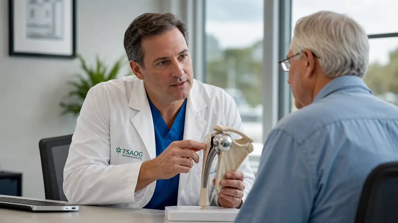
Anatomical total dentures and stemless implants
The anatomical prosthesis faithfully reproduces the biomechanics of a healthy natural joint. It comprises two main components: a metal sphere fixed to the head of the humerus and a concave polyethylene (high-density plastic) surface implanted in the glenoid cavity of the scapula.
For this prosthesis to function optimally, a strict anatomical condition is required: the rotator cuff tendons must be perfectly intact and functional. Without a healthy rotator cuff, the ball is no longer held in place against the glenoid and slips upwards, causing painful impingement and premature loosening of the glenoid component.
On the humeral side, implants have evolved considerably:
- Conventional stem implants : a metal rod (made of titanium or chrome-cobalt alloy) descends into the medullary canal of the arm, ensuring cemented or cementless fixation thanks to a porous osseointegrative coating.
- Stemless prostheses« Designed for patients with good bone density, these implants eliminate the need for a stem to be inserted into the diaphysis. A porous metal anchor or cage is impacted directly into the proximal metaphysis of the humeral head. This approach preserves cortical bone, limits intramedullary bleeding, reduces the risk of intraoperative diaphyseal fracture, and significantly simplifies any future revision surgeries.
What is a reverse shoulder prosthesis and how does it work?
Designed and popularized by Professor Paul Grammont in the late 1980s, the reverse shoulder prosthesis represents a fundamental change in joint geometry. The principle consists of reversing the arrangement of the anatomical parts:
- A metallic hemisphere, called a glenosphere, is securely fixed by a plate screwed to the level of the shoulder blade (the glenoid).
- A concave polyethylene cup is positioned at the top of the humerus, attached to a stem or humeral implant.
This biomechanical inversion shifts the center of rotation of the joint inward and downward. This shift increases the lever arm of the deltoid muscle, the large superficial muscle of the shoulder.
Deprived of its irreparable rotator cuff tendons, the shoulder can no longer center the humeral head in the conventional way. A reverse shoulder prosthesis compensates for this deficit: the deltoid muscle takes over completely to lift the arm without the humeral head retracting under the acromion. This semi-constrained device restores a dramatic active elevation to shoulders that were previously almost entirely unable to move.
Today, the reverse shoulder prosthesis has become the most commonly implanted model. It accounted for 70.4% of shoulder arthroplasties registered in the UK in 2024, and nearly 69% in both Germany and the US, according to the most recent data. [2, 4]. France does not yet have published data on this distribution: the national RENACOT register, supported by SOFCOT, was only created in 2022.
Hemiarthroplasty and resurfacing: partial options
In certain well-defined situations, replacing both articular surfaces is not necessary. This logic of partial implantation, which consists of replacing only the compartment that is actually damaged, is also the one that guides the implantation of a partial knee prosthesis :
- Humeral hemiarthroplasty This procedure involves replacing only the humeral head with a prosthetic sphere, while leaving the native glenoid cartilage intact. It is primarily considered in cases of early avascular necrosis (where the scapular cartilage is not yet damaged) or in certain complex humeral fractures in younger patients. The medium-term drawback remains the risk of secondary erosion of the glenoid cartilage due to friction from the metal head, sometimes requiring further surgery.
- Humeral resurfacing This involves covering the humeral head with a metallic cap without major bone resection or the use of an intramedullary stem. While used in the past to preserve bone in young patients, this technique has seen its indications narrow in favor of stemless anatomical prostheses, which provide more stable fixation and better control of joint stress.
How is the choice decided: age, condition of the rotator cuff, glenoid bone mass
The choice of implant model is determined during the pre-operative consultation based on objective parameters:
| Evaluated parameter | Anatomical prosthesis: | Inverted prosthesis: |
|---|---|---|
| Rotator cuff condition | Intact, healthy, and toned tendons | Massive, irreparable rupture or atrophied muscles |
| Morphology and wear of the glenoid (Walch classification) | Centered glenoid (types A1, A2) or moderate wear | Severe asymmetrical wear (types B2, B3, C) or humeral subluxation |
| Patient's age | Preferred for subjects under 65-70 years old | Very common after age 70; possible earlier in cases of irreparable rupture |
| Underlying diagnosis | Purely centered primary shoulder osteoarthritis | Eccentric glenohumeral osteoarthritis, complex fracture in the elderly, surgical revision |
| Primary muscle engine | Rotator cuff | deltoid muscle |
This tailored assessment ensures a choice adapted to the patient's physical needs and the expected lifespan of the implant.
This consultation should also address the expected results, and not just the technical choice. A reverse prosthesis restores an often spectacular active elevation thanks to the deltoid, but the recovery of the external rotation The outcome remains dependent on the residual condition of the posterior external rotators (infraspinatus and teres minor). When these muscles are already degenerated at the time of surgery, certain movements performed with the elbow close to the body will remain permanently limited: reaching for the ear or phone, accessing a back pocket, turning an ignition key, or grasping a seatbelt. Clearly communicating this limitation before the procedure, rather than discovering it during rehabilitation, largely determines the patient's final satisfaction.
What happens during a shoulder replacement operation?
The surgical procedure to implant a shoulder prosthesis follows a precise, highly standardized technique, where each technical step is meticulously planned. The day's operating procedure unfolds in a manner comparable to other limb arthroplasties, such as the procedure of a hip replacement operation, but the installation position, the approach and the management of the tendons are specific to the shoulder.
Anesthesia, setup and surgical approach
Modern anesthetic protocol favours the combination of loco-regional anesthesia and sedation or light general anesthesia.
Before entering the operating room, the anesthesiologist performs an interscalene block under ultrasound guidance: a local anesthetic is injected around the nerve roots of the brachial plexus in the neck. This block completely numbs sensation in the shoulder and arm for an average of 18 to 24 hours, ensuring a pain-free awakening. In the operating room, intravenous sedation or general anesthesia takes over to ensure the patient's psychological comfort.
The procedure is performed in a seated or semi-seated position, known as the "beach chair" position. The chest is inclined between 45 and 60 degrees, which provides access to the anterior and posterior aspects of the shoulder while reducing cephalic venous pressure and local bleeding.
The most frequently used approach is the deltopectoral pathway. A skin incision of 8 to 12 centimeters is made on the front of the shoulder:
- Access is gained through a natural gap between the pectoralis major muscle and the anterior bundle of the deltoid muscle, by simply separating these muscles without cutting their fibers.
- For an anatomical prosthesis, the tendon of the subscapularis muscle (located in front of the joint) is temporarily cut or detached to open the capsule and expose the glenoid cavity and the humeral head. It will be securely reattached and sutured at the end of the procedure.
- For a reverse prosthesis, the passage can also be made via the deltopectoral route or via the superolateral trans-deltoid route, depending on the surgeon's habits and the conformation of the shoulder.
Implant placement: 3D planning, navigation and customized guides
The precise positioning of prosthetic components is the primary factor in the functional success and long-term survival of the implant. An error of just a few degrees in inclination or retroversion on the glenoid can lead to premature polyethylene wear, instability, or mechanical loosening.
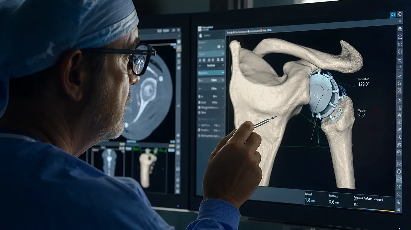
To ensure the reliability of the implantation, modern surgery now incorporates technological assistance tools:
- Preoperative 3D computer planning Based on a previously performed bone scan, a three-dimensional virtual model of the patient's shoulder is created using software. The surgeon simulates the procedure on screen, selects the exact size of the components, determines the depth of bone drilling, and anticipates the ideal axis for implanting the fixation screws.
- Custom cutting guides (PSI) Designed using 3D printing from scanned patient data, these single-use templates adapt perfectly to the bony contours of the glenoid cavity. They guide the surgeon's pilot pin with micrometric precision.
- Surgical navigation and augmented reality Some centers use optical sensors or mixed reality headsets in the operating room. These devices superimpose virtual images of the patient's skeleton and the theoretical implantation axes in real time while the surgeon prepares the bone and inserts the implants. This intraoperative assistance extends a proven approach already used on other joints, particularly during robotic knee replacement.
After reaming the glenoid cavity and resecting the worn cartilage remnant of the humeral head, the trial implants are tested to assess stability, soft tissue tension, and the absence of joint impingement. The final components are then securely anchored (screwed, impacted without cement, or cemented, depending on the chosen technique).
Duration, hospitalization and discharge: from outpatient to traditional inpatient stay
The surgical procedure lasts on average between 45 minutes and 1.5 hours for a first, uncomplicated implantation. A follow-up X-ray is systematically taken immediately after the procedure, in the post-operative monitoring room (recovery room), to verify the correct positioning of the implants.
Hospitalization procedures have changed significantly thanks to enhanced recovery after surgery (ERAS):
- Outpatient hospitalization For independent patients, well supported at home and without serious comorbidities, the procedure can be carried out in a single day. The patient arrives in the morning, is operated on, receives a snack after getting up, then returns home in the late afternoon under the supervision of the home nursing follow-up protocol and local-regional analgesia.
- Short, standard hospital stay This remains the most common approach, with a stay of 24 to 72 hours (1 to 3 nights). This hospitalization allows for monitoring of the initial dressing, adjustment of oral pain management as soon as the anesthetic block wears off, and initiation of the first passive movements supervised by the clinic's physiotherapy team.
Upon discharge, the patient wears a sling or elbow brace to protect the arm during travel, with a prescription for painkillers, sterile dressings and physiotherapy.
The first few weeks: immobility, pain, and daily life
Returning home marks the beginning of a convalescence period where caution and adherence to biomechanical guidelines take precedence over the speed of recovery. The first few weeks play a fundamental protective role: allowing for skin healing, potential tendon attachment, and solid osseointegration of the prosthetic components within the bone.
How long should the splint or abduction cushion be worn?
At the end of the procedure, the operated arm is placed in a splint with the elbow against the body or on a foam abduction cushion keeping the arm slightly away from the torso:
- For a reverse shoulder prosthesis The splint is usually kept in place continuously during 3 to 4 weeks. It acts as a barrier against false reflex movements and prevents the arm from falling backwards.
- For an anatomical prosthesis : the immobilization period is often extended up to 4 to 6 weeks. This increased protection is essential to preserve the surgical reinsertion of the subscapularis tendon, which had to be sectioned and then re-sutured to access the joint. Premature tension could cause the suture to detach.
During this period, the splint should only be removed under specific and safe circumstances: for personal hygiene, dressing, and performing the gentle self-rehabilitation exercises prescribed by the surgeon. The exact date for final removal is determined by the surgeon during the follow-up appointment, and not by the schedule alone.
How to sleep after shoulder surgery?
Sleep is among the primary concerns of patients. Finding a restful and pain-free position requires adapting one's nighttime setup:
- The semi-seated position on the back (reference position) For the first two to three weeks, sleeping with your torso slightly inclined using several pillows or a triangular cushion significantly reduces capsular and muscular tension in the shoulder. It is essential to place a cushion, a thin pillow, or a rolled-up towel under the elbow of the operated arm: this support prevents the elbow from falling backward toward the mattress (retropulsion movement), which would put sudden strain on the front of the joint.
- Side sleeping not operated If lying on your back proves difficult, you can lie on your unaffected side. In this case, place a bolster or large pillow across your chest: the operated arm will rest gently on it, preventing it from collapsing forward or rotating excessively.
- The formal ban Sleeping on the operated side is not recommended for at least 3 to 4 months. Direct pressure of body weight on the implant would cause acute pain and compromise the stability of the still young bone fixations.
Pain management and scar care
After the interscalene anesthetic block wears off (between 18 and 24 hours post-surgery), pain can intensify if not anticipated. The pain management protocol relies on the systematic and regular intake of prescribed medications (paracetamol, anti-inflammatories as directed by a doctor, and mild level 2 opioids for the first few days), without waiting for the pain to develop.
Cryotherapy (application of cold) represents an indispensable local weapon: applying an ice pack or a thermal cushion wrapped in a clean cloth to the shoulder for 20 minutes, 3 to 4 times a day, provides a powerful analgesic and decongestant effect.
Regarding the scar, nursing care is provided every 2 to 3 days at home with a sterile dressing. Waterproof occlusive dressings are not recommended initially, as they promote maceration and increase the risk of local skin infection. Surgical staples or non-absorbable sutures are generally removed around the fifteenth postoperative day. Once the scar is completely closed and dry, gentle massages with a moisturizing or healing cream help soften the tissues and prevent adhesions.
Washing, dressing, eating: forbidden actions and life with one arm
Adapting to everyday movements requires some ergonomic tips to protect the operated shoulder:
- For the dressing Opt for loose-fitting clothing with button or zipper openings at the front rather than tight-fitting sweaters that have to be pulled over the head. The essential rule is to always put on the sleeve on the operated side first, then the unoperated arm. To undress, do the reverse: first remove the sleeve on the unoperated side, then gently free the operated arm without lifting it away from the body.
- For the toilet : washing the operated arm and axillary hollow is done by leaning slightly forward, torso inclined, letting the arm hang by gravity (pendulum position): this naturally detaches the armpit from the thorax without active muscular exertion.
- For meals and keyboard typing : with the elbow resting on an armrest or on the table, active use of the hand, fingers and wrist is permitted and encouraged from the first days (holding a book, writing, using a fork, handling a computer mouse).
- Strictly prohibited actions :
- Carrying the slightest load with the operated arm (no lifting a pack of water, a suitcase or a saucepan).
- Use the operated hand or elbow for support when getting up from a chair or bed.
- Extend your arm behind your body (retropulsion) or bring your hand behind your back.
- In the case of an anatomical prosthesis: force external rotation (arm turned outwards) in order not to tear the freshly repaired subscapular tendon.
- In the case of a reverse prosthesis: combine adduction, extension and forced internal rotation (the gesture of putting on a jacket by throwing the arm back), which constitutes the main position of dislocation of the implant.
These instructions follow the same logic of protection as movements prohibited after hip replacement surgery : each implant has its own specific point of weakness, and the key to preventing early complications is learning to recognize it in order to avoid it on a daily basis.
Rehabilitation and return to activities: the timeframes to know
Functional rehabilitation is the cornerstone of successful shoulder arthroplasty. A prosthesis that is perfectly implanted radiologically will only provide optimal functional results if the physiotherapy treatment strictly adheres to the physiological healing and muscle strengthening timelines.

Phase-by-phase rehabilitation protocol (summary table from day 0 to 1 year)
Rehabilitation after shoulder replacement does not aim for immediate performance: it progresses in stages, following the physiological timelines of tendon healing and osseointegration. Four phases follow one another, defined by the type of mobilization permitted. passive mobilization This means that the physiotherapist mobilizes the arm without any contraction from the patient. active mobilization allows the patient to raise their arm independently using their unaffected arm, a stick, or a pulley system. Mobilization active free, and then reinforcement against resistance, only come afterwards.
The target of the reinforcement differs depending on the implant placed: on an anatomical prosthesis, the work focuses on the centering ensured by the rotator cuff; on a reverse shoulder prosthesis, it targets the deltoid muscle and the scapular fixators, which have become the sole motors of elevation.
| Phase and period | Main objectives | Types of exercises allowed | Restrictions and prohibitions |
|---|---|---|---|
| Phase 1 Day 0 to 4-6 weeks | Scar protection, pain relief, stiffness prevention | Gentle passive mobilization, pendulum work, finger, wrist and elbow movements | Continuous splint wear; no active elevation; no load carrying |
| Phase 2 End of immobilization in the 3rd month | Weaning from the splint with surgical approval, gentle increase in range of motion | Assisted active mobilization (stick, pulley therapy), hot water hydrotherapy | Weaning at 4 weeks for a reversed prosthesis, 6 weeks for an anatomical one; no sudden movements; no weight-bearing on the operated arm |
| Phase 3 Months 3 to 4 | Recovery of active elevation and daily movements | Free active movements, self-rehabilitation, scapular postural work | Loads limited to 1-2 kg; no strong movements |
| Phase 3 Months 4 to 6 | Targeted muscle strengthening according to the implant, joint stability | Low-resistance elastic bands, gentle support, maintenance stretching | Avoid repetitive movements above the horizontal while carrying a load |
| Phase 4 Months 6 to 12 | Functional consolidation, endurance and maximum autonomy | Endurance training, spatial coordination, resumption of adapted leisure activities | Contact sports, combat sports and violent throwing sports are prohibited. |
When can you drive after shoulder surgery?
Resuming driving requires full recovery of emergency reflexes, sufficient grip strength, and the ability to turn the steering wheel with both hands instantly and without pain.
In practice, Driving is possible between the 2nd and 3rd postoperative months (usually around 8 to 10 weeks).
This resumption of activity depends on strict clinical criteria:
- The protective splint must be completely removed during the day.
- Active lifting and arm control should allow the steering wheel to be maneuvered without locking or sharp pain in case of sudden avoidance.
- The use of strong painkillers that can impair alertness must have ceased.
This 2-3 month timeframe remains a functional average, and not an automatic authorization. The formal decision regarding resuming driving rests with the surgeon., during the 2nd or 3rd month check-up. He specifically checks the restoration of the avoidance reflex The ability to make a wide and rapid steering turn with both hands, without apprehension, joint locking, or pain. Resuming driving before this agreement exposes you not only to the risk of an accident due to an impossible emergency maneuver, but also to a dispute of coverage by the insurer in the event of an accident occurring during the period of incapacity.
Once the agreement is obtained, it is recommended to start with short, familiar journeys on quiet roads before tackling heavy traffic or long motorway journeys.
Sport and carrying loads: what is becoming possible again
Returning to leisure activities is considered on a highly individualized basis, depending on the type of prosthesis, the regained range of motion, and the sport practiced. Returning to work, which is subject to different criteria, is detailed further down in the section on sick leave.
- Authorized and encouraged sporting activities From the second month onwards, brisk walking, hiking on stable terrain, and stationary cycling are recommended. Gentle swimming (breaststroke without exertion) and road cycling on flat terrain are generally permitted around the third or fourth month. Golf (gradually resuming the swing), gentle gymnastics, or light bowling can be considered between four and six months after a medical check-up.
- Activities that are discouraged or prohibited To preserve the anchoring of the metal parts and prevent wear on the high-density plastic insert, contact sports (rugby, judo, football) and violent racket sports (competitive tennis, squash) are strongly discouraged in the long term. The same applies to the repeated wear Heavy loads exceeding 10 to 15 kilograms, especially above shoulder level, should be avoided. However, occasional light lifting remains possible once recovery is complete, according to the limits detailed at the end of this article.

Results, prosthesis lifespan and complications
Shoulder replacement surgery positively transforms patients' daily lives by eliminating chronic pain that has often been present for years. However, like any major reconstructive surgery, it has biomechanical limitations and specific risks that must be discussed openly and transparently.
Mobility and function restored, including the limit in external rotation after reverse prosthesis
The analgesic effect of the intervention is its most consistent and predictable outcome: published series report complete or near-complete pain relief in 68% to 91% of patients [9], and overall satisfaction after reverse prosthesis is between 78 % and 88 % [7]. International functional scores (Constant score and ASES score) gain on average 35 to 45 points, as measured in the medium and long term in available systematic reviews. [7] No solid data documents this gain in the first year. This analgesic reliability characterizes arthroplasty of large joints in general, as shown by the data published on the success rate of a knee replacement.
The increase in range of motion varies depending on the prosthetic model implanted:
- With a total anatomical prosthesis When the rotator cuff is healthy, the joint regains near-normal kinematics. Active anterior elevation reaches an average of nearly 150 degrees at long retraction, compared to approximately 119 degrees before surgery. [10], with good preservation of internal and external rotations.
- With a reverse shoulder prosthesis : recovery of active anterior elevation is very satisfactory, reaching an average of 126 degrees for a gain of about fifty degrees [7]. The patient can easily bring their hand to their mouth, the back of their neck, and the top of their head without tilting their head. However, the active external rotation The ability to extend the forearm outward with the elbow at the side often remains limited, as should have been discussed during the preoperative consultation. This biomechanical restriction is due to prior degeneration of the posterior external rotators (infraspinatus and teres minor): it is therefore determined by the patient's preoperative muscle condition, and not by the quality of the surgical technique. The same studies also place the average external rotation at only around 22 degrees. [7]. To overcome this deficiency, modern implants use lateralized glenospheres or the BIO-RSA autologous bone grafting technique, which improve angular range of motion.
Implant longevity, radiological follow-up, and prosthesis revision
Advances in metallurgy and developments in highly cross-linked polyethylene have significantly increased the durability of implants. National registries place survival at around 94 to 96 % at 10 years: the British registry reports a cumulative risk of revision of 6.1 % for the anatomical prosthesis and 4.1 % for the reverse prosthesis at this time. [2], and the Norwegian 94 % survival registry of inverteds [3]. Beyond 15 years, data remains scarce and heterogeneous: a historical series reports 88 % at 15 years and 85 % at 20 years [11], a recent series 72 % at 20 years [10].
This longevity depends closely on the accuracy of the initial positioning of the glenoid, the quality of the recipient bone, and the level of mechanical stress imposed by the patient.
A protocol of regular radiological follow-up is essential, even in the total absence of pain:
- Follow-up appointments are scheduled at the first month, the third month, and then one year after the intervention.
- Beyond that, a check-up visit with frontal and lateral radiographs every 2 to 3 years allows monitoring for the absence of wear lines or silent peri-prosthetic osteolysis.
In cases of aseptic loosening or advanced wear of the plastic insert, revision surgery is possible. For a failing anatomical prosthesis, conversion to a reverse prosthesis provides excellent functional results. These second-stage procedures follow the same principles as...’a hip replacement revision : they are technically more demanding than the initial placement, require anticipating the loss of bone capital and justify even more thorough preoperative planning.
Possible complications: infection, dislocation, loosening, scapular notch, stiffness, nerve damage, complex regional pain syndrome (CRPS)
In a multicenter series of over 6,000 patients, the overall complication rate reached 10.7% after anatomical prosthesis and 8.9% after reverse prosthesis, with a surgical revision rate of 5.6% and 2.5% respectively. [6]. These rates climb sharply in revision surgery, where specialist journals report up to 40% of complications compared to approximately 15% in primary surgery. [5]. All candidates for intervention must be informed of the potential risks:
- Periprosthetic infection (in the order of 1 to 2 % in the national registers) [3], (more so in long-term follow-up series and revision surgery): a serious complication that may require surgical joint lavage or complete implant replacement in one or two stages under targeted antibiotic therapy. The shoulder has a specific sebaceous skin flora dominated by Cutibacterium acnes, a slow-growing anaerobic bacterium that requires meticulous antiseptic preparation and strict antibiotic prophylaxis protocols in the operating room.
- Prosthetic dislocation (approximately 2.5 % in total, 1 to 5 % for the first-line reversed [12]): it is the primary cause of revision surgery for reverse prosthesis [2] and occurs during movements combining extension and forced internal rotation. It requires reduction under anesthesia, sometimes followed by a short period of immobilization or implant adjustment if instability recurs. This prosthetic instability should not be confused with chronic shoulder instability in the native joint, which concerns much younger patients and involves a completely different stabilization surgery.
- Glenoid loosening It mainly concerns the polyethylene cup of anatomical prostheses, which is subjected to eccentric shear stresses (rocking-horse effect). Two often confused realities must be distinguished: loosening radiological, visible on approximately 15 % of the all-polyethylene cemented glenoids, and the unsealing symptomatic leading to surgical revision, which affects approximately 6 % of cases [13]. 3D planning assistance significantly reduces this phenomenon today.
- The scapular notch Specific to reverse shoulder arthroplasty, this erosion of the inferior neck of the scapula is caused by repeated contact of the humeral cup against the bone during arm adduction. Its incidence varies from 10⁻¹TP³T to over 60⁻¹TP³T depending on the implant design and the positioning of the glenosphere. [7] It reached up to 70 % on historical medialized models, compared to approximately 23 % on lateralized designs. It most often remains asymptomatic, and its occurrence is now limited by the lowering and lateralization of the new glenospheres.
- Postoperative stiffness and algodystrophy Stiffness can occur if rehabilitation is poorly balanced. Complex regional pain syndrome (algodystrophy) combines edema, hypersensitivity and stiffness lasting several months, requiring specific medical management and the temporary cessation of painful physiotherapy maneuvers.
- Nerve damage (approximately 1.3 % in primary surgery, of which 0.64 % for the axillary nerve alone) [8]The axillary nerve and the trunks of the brachial plexus run in the immediate vicinity of the surgical site. Intraoperative traction can cause paresis or numbness of the arm, which is almost always transient (neurapraxia resolving within a few weeks or months).
Price, reimbursement and work stoppage
Anticipating administrative, financial, and professional procedures is an integral part of preparing for shoulder surgery. Clear information about costs and recovery time allows you to organize your convalescence under the best possible conditions.
Cost of the intervention and coverage (sector, overruns, long-term illness, health insurance)
In France, shoulder replacement surgery is a coded surgical procedure in the Common Classification of Medical Procedures (CCAM), specifically chapter 13.3.2.6 dedicated to scapulohumeral arthroplasty. The main code is MEKA006 (scapulohumeral joint replacement by total prosthesis), the basic rate of which in sector 1 amounts to 519.52 euros as of January 1, 2026. Code MEKA008, which covers the same procedure combined with rotator cuff repair, is priced at 598.59 euros.
Two points are important here. First, The CCAM (French Classification of Medical Procedures) does not distinguish between anatomical and reverse prostheses. Both procedures fall under the same codes. Furthermore, this fee only covers the surgical procedure itself. The implant is covered separately under the List of Products and Services (LPP). Hospital stays and prosthetic materials are covered by Health Insurance based on the standard reference rates.
The financial elements are divided into several categories:
- The practitioner's area of practice :
- In sector 1 (or in a public hospital without private practice), no additional fees are charged; care is reimbursed at the Social Security rate.
- In sector 2 with unrestricted fees (private clinics or private consultations at the hospital), additional fees may be charged by the surgeon and the anesthesiologist. A personalized and detailed written estimate must be provided to the patient before the procedure.
- The role of supplementary health insurance (mutual insurance) : reimbursement of additional fees, daily hospital allowance and comfort options (private room, television) depends directly on the guarantees subscribed to in the mutual insurance contract.
- The Long-Term Illness (ALD) scheme : if the intervention is motivated by a recognized systemic pathology in ALD (such as severe rheumatoid arthritis or spondyloarthritis), the exemption from the co-payment applies to 100 % on the Social Security reimbursement bases [1]. The patient is responsible for excess fees, the fixed co-payment, the medical deductible, and the hospital daily fee. Be careful not to misunderstand: Isolated shoulder osteoarthritis does not entitle the patient to long-term illness benefits.. This scheme only applies if the patient is also recognized as having a long-term illness that qualifies for exemption from other medical conditions.
No standard amount can be announced in advance. The additional fees charged by the surgeon and anesthesiologist vary considerably depending on the practitioner, the facility, and the region. The written estimate provided before the procedure is therefore the only legally binding document and the only reliable basis for calculating the remaining balance. It must be sent to the supplemental health insurance provider several weeks before hospitalization: this timeframe allows for written confirmation of coverage from the insurer and avoids any unpleasant surprises when the bill arrives.
Duration of sick leave depending on the occupation and return to work
The doctor issues the sick leave prescription upon discharge from the clinic. Its duration varies considerably depending on the physical strain placed on the upper limb during work activity:
- For sedentary and office jobs : computer work, secretarial work or tertiary activities generally allow for a return between 6 and 8 weeks post-operative. If the daily commute does not require driving and teleworking is feasible, an early return to work can be discussed.
- For intermediate activities involving frequent travel Commercial or supervisory professions require an average waiting period of 2 to 3 months, corresponding to the time needed to resume driving safely and with the surgeon's approval.
- For manual laborers and those in physically demanding jobs For tradespeople, construction professionals, farmers, or healthcare workers handling patients, sick leave usually extends from 4 to 6 months.
A pre-return visit A consultation with the occupational physician must be scheduled before the end of the sick leave. This consultation allows for planning ahead for workplace adjustments, a temporary exemption from heavy lifting, or the implementation of a therapeutic part-time schedule (half-time work) to ensure a safe return to work without jeopardizing the future of the prosthesis.
Frequently Asked Questions
Is it possible to have an MRI scan with a shoulder prosthesis?
Yes. Modern shoulder prostheses are made from titanium, chromium-cobalt, and high-density polyethylene alloys—non-ferromagnetic materials. There is no risk of detachment or dangerous overheating due to a magnetic field.
However, it is essential to inform the radiology team of the presence of the prosthesis before the examination. The metallic components cause an artifact (shadow area or image distortion) in the surgical area. Radiologists then adapt their acquisition sequences (MARS sequences for reducing metallic artifacts) to obtain precise images, whether the examination concerns the operated shoulder or another part of the body.
Does the prosthesis trigger airport security gates?
Sometimes. Depending on the sensitivity of the airport's metal detectors and the metallic mass of the implant, the metal detector may sound as you pass through, without this causing any particular difficulty.
You will receive an implant card or surgical certificate confirming the placement of your prosthesis upon your discharge from the clinic. You can keep it in your wallet when traveling. At airports equipped with millimeter-wave body scanners, the prosthetic component will appear clearly on the screen, simplifying the security check.
From what point can I sleep on the side I operated on?
It is advisable to wait at least 3 to 4 months before considering sleeping directly on the operated shoulder.
Direct pressure from the body's weight on the joint compresses the bursa, the remodeling muscle masses, and the still-developing bone structures. Even after three months, this position should be resumed very gradually: if discomfort or a dull ache develops during the night, this habit should be postponed for a few more weeks.
Is it possible to have surgery on both shoulders?
Yes, but never during the same procedure. Primary glenohumeral osteoarthritis and bilateral rotator cuff tears are common in orthopedic consultations, and it is perfectly possible to operate on both shoulders successively.
Simultaneous surgery on both sides under the same anesthesia is, however, strictly avoided. Mandatory immobilization of an upper limb with a splint would deprive the patient of all vital autonomy (inability to eat, wash, or dress independently). A waiting period of 6 months to 1 year is usually respected between the two surgeries: this period of time allows the first shoulder to have recovered sufficient strength, mobility and stability to perform daily activities during the convalescence of the second.
How much weight can one bear in the long term?
A joint prosthesis is not indestructible: the mechanical stresses applied to the arm are transmitted directly to the anchoring zone of the implant in the bone of the glenoid and humerus.
Once muscle consolidation and strengthening are achieved (around 6 to 12 months), carrying loads occasional Carrying a bag weighing between 5 and 10 kilograms is perfectly acceptable for everyday activities (shopping bags, household chores, light luggage). However, lifting repeated loads exceeding 10 to 15 kilograms, the practice of heavy weight training with bench press or jobs requiring carrying loads above shoulder level are strictly prohibited to prevent premature aseptic loosening.
Is it normal to feel or hear the prosthesis?
Yes, this sensation is common and harmless. During the first few months, some patients may experience a slight metallic clicking or a soft rubbing noise during certain rotational movements: this is simply the regular contact between the metal surface and the polyethylene dome, or scar tissue sliding around the joint.
Similarly, a temporary sensation of a "heavier" limb or anterior muscle tension is common while the deltoid muscle adapts to its new working length. As long as these sounds or sensations are not accompanied by sharp pain, sudden locking, or signs of local inflammation, they are normal and will subside with the course of rehabilitation.
Successfully complete your recovery with personalized support
The timeframes and protocols presented in this guide represent averages observed in patients who underwent surgery for a shoulder prosthesis. However, each treatment path remains unique. Your recovery speed depends closely on specific parameters: the initial state of your rotator cuff tendons, the quality of your glenoid bone stock, the type of implant chosen (anatomical model or reverse shoulder prosthesis) as well as your regularity in self-rehabilitation exercises.
To navigate each stage smoothly—from gradually weaning yourself off the brace to resuming driving and your leisure activities—medical expertise remains essential. Only regular clinical and radiological monitoring allows for proper osseointegration of the implants, prevents stiffness, and enables the physiotherapist to adjust their work according to your priorities, whether it's raising your arm again without pain or resuming appropriate physical activity.
If you suffer from persistent shoulder pain or are considering surgery, I encourage you to schedule a consultation appointment. We will conduct a complete assessment of your joint in order to determine the most appropriate surgical and rehabilitative solution for your situation.
Sources
The numerical data in this article are derived from national arthroplasty registries and indexed systematic reviews, referenced below. Complication and survival rates vary considerably depending on the implant design, the surgical indication, and the duration of follow-up: the ranges presented reflect this real heterogeneity, and not an inaccuracy in the interpretation.
- Service-Public.gouv.fr (DILA), Coverage of a long-term illness (ALD) by Health Insurance, Page verified on December 18, 2025. service-public.gouv.fr
- National Joint Registry, The National Joint Registry 22nd Annual Report, 2025. ncbi.nlm.nih.gov
- Hole RM, Fenstad AM, Gjertsen JE, Hallan G, Veurne ON. Influence of design features and brand of reverse shoulder arthroplasties on survivorship and reasons for revision surgery: results of 5,494 arthroplasties reported to the Norwegian Arthroplasty Register 2007-2022. Acta Orthopedica, 2024;95:463-471. pmc.ncbi.nlm.nih.gov
- Rupani N, Combescure C, Silman A, Lübbeke A, Rees J. International trends in shoulder replacement: a meta-analysis from 11 public joint registers. Acta Orthopedica, 2024;95:348-357. pmc.ncbi.nlm.nih.gov
- Barco R, Savvidou OD, Sperling JW, Sanchez-Sotelo J, Cofield RH. Complications in reverse shoulder arthroplasty. EFORT Open Reviews, 2016;1:72-80. pubmed.ncbi.nlm.nih.gov
- Parada SA, Flurin PH, Wright TW, Zuckerman JD, Elwell JA, Roche CP, Friedman RJ. Comparison of complication types and rates associated with anatomic and reverse total shoulder arthroplasty. Journal of Shoulder and Elbow Surgery, 2021;30(4):811-818. pubmed.ncbi.nlm.nih.gov
- Doyle TR, Downey S, Hurley ET, Klifto C, Mullett H, Denard PJ, Garrigues GE, Menendez ME. Midterm outcomes of primary reverse shoulder arthroplasty: a systematic review of studies with minimum 5-year follow-up. JSES Reviews, Reports and Techniques, 2023;4(1):1-7. pmc.ncbi.nlm.nih.gov
- North D, Hones KM, Jenkins P, Sipavicius E, Zermeño Salinas JL, Hao KA, Schoch BS, Wright TW, Gulotta LV, King JJ. How common is nerve injury after reverse shoulder arthroplasty? A systematic review. Journal of Shoulder and Elbow Surgery, 2023;32(4):872-884. pubmed.ncbi.nlm.nih.gov
- Ricchetti ET, Abboud JA, Kuntz AF, Ramsey ML, Glaser DL, Williams GR Jr. Total shoulder arthroplasty in older patients: increased perioperative morbidity? Clinical Orthopedics and Related Research, 2011;469(4):1042-1049. pmc.ncbi.nlm.nih.gov
- Patel AV et al. Anatomic total shoulder arthroplasty: long-term clinical, radiographic, and patient-reported outcomes. Journal of the American Academy of Orthopedic Surgeons, 2026;34(16):e2226-e2232. pubmed.ncbi.nlm.nih.gov
- Deshmukh AV, Koris M, Zurakowski D, Thornhill TS. Total shoulder arthroplasty: long-term survivorship, functional outcome, and quality of life. Journal of Shoulder and Elbow Surgery, 2005;14(5):471-479. pubmed.ncbi.nlm.nih.gov
- Olson JJ, Galetta MD, Keller RE, Oh LS, O'Donnell EA. Systematic review of prevalence, risk factors, and management of instability following reverse shoulder arthroplasty. JSES Reviews, Reports and Techniques, 2022;2(3):261-268. pmc.ncbi.nlm.nih.gov
- Kim DM, Aldeghaither M, Alabdullatif F et al. Loosening and revision rates after total shoulder arthroplasty: a systematic review of cemented all-polyethylene glenoid and three modern designs of metal-backed glenoid. BMC Musculoskeletal Disorders, 2020;21:114. pmc.ncbi.nlm.nih.gov
This article was written and medically reviewed by Dr. Frédéric Sailhan. The numerical data are from national arthroplasty registries and systematic reviews listed in the sources, accessed in September 2026.
This article is for informational purposes only and is not a substitute for medical advice. The timeframes, thresholds, and percentages mentioned are approximate and based on common practice: only your surgeon can determine the specific guidelines applicable to your prosthesis, your anatomy, and your activity level.

 English
  French